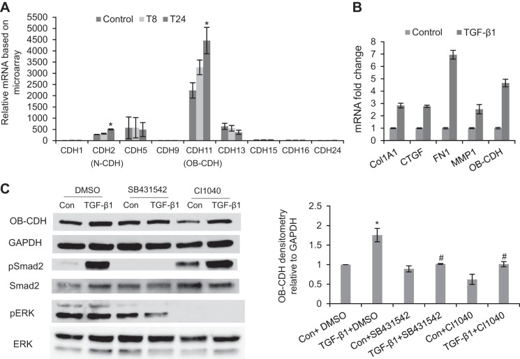
Figure 3.
TGF-β1 treatment up-regulates expression of CDH11 in VICs through both the Smad2 and the ERK signaling pathways. A) Normalized expression value of different cadherins in VICs detected by microarrays. CDH2, CDH5, CDH11, and CDH13 were the abundantly expressed cadherins in VICs. However, only CDH2 and CDH11 were significantly up-regulated after 24 h of TGF-β1 treatment. T8, TGF-β1 treatment for 8 h; T24, TGF-β1 treatment for 24 h. *P < 0.05 vs. control. B) Confirmed by qRT-PCR, TGF-β1 treatment for 24 h increased mRNA level of CDH11 and also up-regulated expression of the fibrogenic genes, including Col1A1, CTGF, FN1, and MMP1. This is representative of ≥3 independent qRT-PCR experiments. Error bars = sd. C) At the protein level, TGF-β1 treatment (24 h), which activated pSmad2, also up-regulated CDH11 protein expression. The effect of TGF-β1 on CDH11 expression was blocked by inhibiting either Smad2/3 signaling via SB431542 or ERK signaling via CI1040. Con, control (untreated), *P < 0.05 vs. Con + DMSO; #P < 0.05 vs. TGF-β1 + DMSO.

