Figure 2.
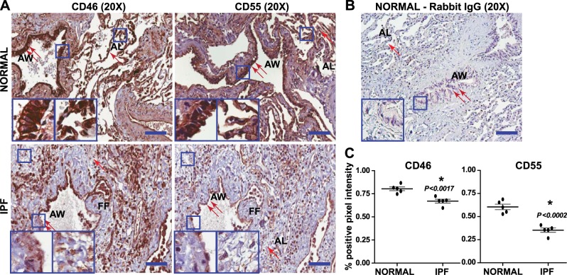
Loss of CD46 and CD55 in IPF lungs. Comparative immunohistochemical analysis of paraffin-embedded human IPF lung biopsy explants obtained at lung transplant and tissue resected from normal (non-IPF) lungs was conducted with CD46 and CD55 staining. A) Top panels: normal lungs at the time of resection for other diseases. Normal lung architecture with significant CD46 and CD55 expression in the airway (2 arrows) and the alveolar epithelium (1 arrow). Bottom panels: IPF lung tissue biopsy. Disrupted airway epithelium (2 arrows) and alveolar epithelium (1 arrow), with loss of CD46 and CD55 staining (DAB, brown) appearing in the mesenchymal cells at the fibroblastic foci (FF). Nuclei were counterstained with hematoxylin (blue). Insets: airway (left panels); alveoli and interstitium (right panels). Representative lesions observed from 5 different patients are presented. AW, airway; AL, alveoli. Scale bars = 100 μm (×20). B) Normal lungs immunostained with corresponding rabbit IgG. Scale bars = 100 μm (×20). C) Intensity analysis of CD46 and CD55 staining in 5 normal and 5 IPF tissue sections. Values represent means ± sem; unpaired t test. *P < 0.05.

