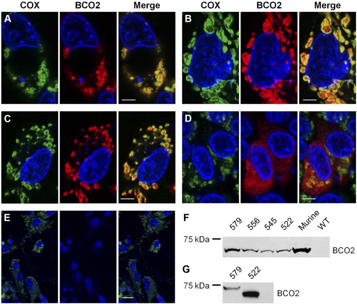
Figure 3.
Human BCO2 localization is dependent on an N-terminal mitochondrial targeting sequence. A–D) HepG2 cells were transfected with expression plasmids encoding the 579-aa (A), 556-aa (B), 545-aa (C), and 522-aa (D) isoforms of human BCO2. E) As a control, untransfected HepG2 cells were used. Immunostaining was performed with anti-V5 antibody for BCO2 (red) and anti-COX IV antibody (green). DAPI was used to stain the nuclei. Merged images show a colocalization of BCO2 and COX IV for the 579-, 556-, and 545-aa BCO2 isoforms. Cells expressing the 522-aa isoform do not exhibit such staining. Untransfected control cells display no anti-V5 antibody staining. Scale bars = 5 μm (A–D); 25 μm (E). F, G) Immunoblots for BCO2 from transfected HepG2 cells (F) and transformed E. coli (G). As a control, HepG2 cells that express murine BCO2 and untransfected HepG2 (WT) were used. Total protein (20 μg) was separated by SDS-PAGE. Recombinant BCO2 was stained with anti-V5 antibody.

