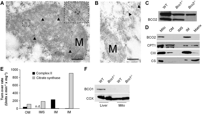
Figure 5.
BCO2 localizes to the inner membrane of the mitochondria. A, B) COS7 cells were transfected with the 545-aa (A) and the 522-aa (B) BCO2 isoforms. Immunogold labeling was performed with anti-V5 antibody and imaged with transmission electron microscopy. Immunogold labeling for the human 545-aa isoform was found at the inner membrane of mitochondria. Cells transfected with the 522-aa BCO2 isoform had no observable gold particles within the mitochondria. Arrows indicate gold particles; M, mitochondria. Scale bars = 200 μm. Inset: zoom of the marked area. Note the gold particles associate with the inner mitochondrial membrane. Several images were taken, representative samples are shown. C) Immunoblot analysis for BCO2 of whole liver protein extracts of WT, Bco1−/−, and Bco2 mice confirmed the specificity of the BCO2 antiserum. Note that the antibody shows a nonspecific cross-reaction with a protein of ∼80 kDa in size. D) Hepatic mitochondria were isolated and fractionated. Protein extracts from total mitochondria (Mito), outer membrane (OM), inter membrane space (IMS), inner membrane (IM), and matrix were subjected to immunoblot analysis for BCO2, carnitine palmitoyltransferase 1 (CPT1) and complex III (CIII). E) Test for enzymatic activity of citrate synthase (matrix) and complex II (succinate dehydrogenase, inner membrane) of different mitochondrial fractions. n.d., not detectable. F) Immunoblot analysis for BCO1 with protein extracts of whole liver and isolated mitochondria of WT and Bco1−/− mice. Analysis in Bco1−/− mice confirmed the specificity of the BCO1 antiserum.

