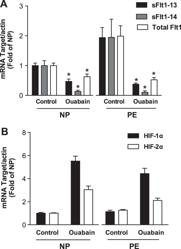
Figure 4.

mRNA expression studies for sFlt1 and HIF-1α. mRNA levels were normalized to actin and presented as ratios relative to the normal explants. A) Quantitative real-time PCR showing reduction in mRNA for total Flt1, sFlt1-13, and sFlt1-14 isoforms in normal (n=6) and preeclamptic (n=6) villous explants exposed to 500 nM ouabain. B) Quantitative PCR shows an increase in HIF-1α and HIF-2α mRNA in normal (n=6) and preeclamptic (n=6) explants exposed to 500 nM ouabain. NP, normal pregnancy; PE, preeclampsia. *P < 0.05 vs. control; Student's t test.
