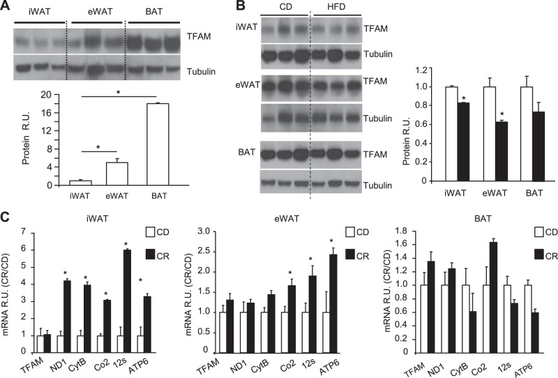
Figure 1.
TFAM expression in WAT and BAT from mice fed HFD or subjected to CR. A) Top panel: TFAM expression was analyzed by Western blot analysis of iWAT, eWAT, and BAT. Bottom panel: quantitation of data (n=6 samples/tissue). *P < 0.05. B) Left panel; TFAM expression was analyzed by Western blot in iWAT, eWAT, and BAT from mice fed a CD or HFD at 16 wk of age. Data are representative of 6 samples/genotype. Right panel: quantitation of data. *P < 0.05. C) TFAM and mRNA levels of mitochondria-encoded genes in mice fed a CD or subjected to CR, using qPCR of iWAT, eWAT, and BAT from 16-wk-old male mice. Data are means ± sem of 6 samples/group. *P < 0.05.

