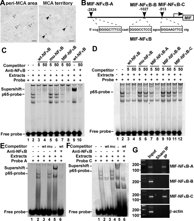
Figure 2.
Identification of the functional NFκB binding sites on the human MIF promoter. A) Brain slices from BALB/c mice subjected to 2 h tMCAl followed by 2 h reperfusion were stained for NFκB p65. In the MCA territory, p65 exhibited nuclear localization (solid arrowheads), while in the cortical peri-MCA region, p65 diffused in the cytoplasm (open arrowheads). Scale bars = 20 μm. B) Schematic diagram shows the relative location of the putative NFκB binding sites on the human MIF promoter, and the corresponding sequence of the oligonucleotides. C, D) GSAs were performed using a wt-NFκB consensus oligonucleotide labeled with IRDye 700 as the probe. Lane 1 is labeled probe alone without nuclear extract. Incubation of the wt-NFκB probe with nuclear extract resulted in a shifted band composed of DNA-protein complex (lane 2). Addition of anti-NFκB p65 monoclonal antibody in the incubation mixture as in lane 2 resulted in a supershifted band (C, lane 9). Competition assays were performed by adding 5- or 50-fold molar excess of unlabeled wt-NFκB oligonucleotide (lanes 3, 4), mu-NFκB oligonucleotide (lanes 5, 6), MIF-NFκB (C, lanes 7, 8), MIF-NFκB-A (D, lanes 7, 8), MIF-NFκB-B (D, lanes 9, 10), MIF-NFκB-C (D, lanes 11, 12), and 50-fold molar excess of wt-NFκB (C, lane 10). E, F) GSAs and supershift assays were performed using MIF-NFκB-A oligonucleotide (E) or MIF-NFκB-C oligonucleotide (F) labeled with IRDye 700 as probes. Lane 1 is labeled probe alone without nuclear extract. Incubation of IRDye 700-MIF-NFκB-A or -C with nuclear extracts resulted in a shifted band (lane 2). Addition of anti-NFκB p65 monoclonal antibody resulted in a supershifted band (lane 5). Competition assays were achieved by adding 100-fold molar excess of unlabeled wt-NFκB (lanes 3, 6) or the same amount of mu-NFκB (lane 4). G) ChIP assay; details are described in Materials and Methods. Monoclonal rabbit-anti-p65 antibody was used to precipitate DNA fragments that could bind to NFκB p65. Normal rabbit serum was used for sham IP control. Target DNA fragments containing sequences corresponding to MIF-NFκB-A, -B, and -C were amplified by PCR. PCR products were resolved on 1.5% agarose gel. β-Actin was amplified as control.

