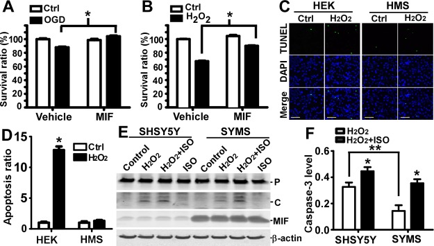
Figure 4.

MIF protected neurons by inhibiting caspase-3 activation. Cortical neurons cultured from E14 mouse embryos were subjected to an in vitro model of ischemia/reperfusion or oxidative stress. A) In vitro ischemia/reperfusion was achieved by challenging the primary neuronal culture in glucose-free medium under hypoxic conditions for 2 h (ischemia period), followed by 16 h incubation under normoxic conditions after medium exchange to normal medium (reperfusion period). Purified human his-tagged MIF (200 ng/ml) or vehicle was added in the medium during the entire experiment. Cell viability was assessed by using MTS assays. Survival ratio represents the average absorbance to control absorbance. Values represent means ± sd, n = 10. *P < 0.01; Student's t test. B) Oxidative stress was induced by 50 μM of H2O2 for 16 h in normal medium. Purified human his-tagged MIF (200 ng/ml) or vehicle was added in the medium during the entire experiment. Cell viability was assessed by using MTS assays. Survival ratio represents the average absorbance to control absorbance. Values represent means ± sd, n = 6, *P < 0.001; Student's t test. C) HEK293 and MIF stable HMS cells were treated with 50 μM H2O2 for 24 h in medium lacking sodium pyruvate, followed by TUNEL assay (green channel). DAPI (blue channel) was used to stain nuclei. Scale bars = 100 μm. D) Quantification of cell numbers identified by DAPI staining and apoptotic cells identified by TUNEL labeling by ImageJ. Signals were averaged from 5 randomly selected views. Values represent means ± sd, n = 3. *P < 0.01 vs. cells without H2O2 treatment; 2-way ANOVA with Bonferroni posttests. E) SHSY5Y and MIF stable SYMS cells were treated with 200 μM H2O2 for 16 h. MIF inhibitor ISO-1 (50 μM) was added to block the effect of MIF. Cells were lysed by Chap cell extract buffer. Lysate was resolved on a 12% Tris-tricine SDS-PAGE gel, and protein levels were analyzed by Western blot. An anti-caspase-3 antibody recognizing both pro- and cleaved caspase-3 was used. F) Quantification of E. Ratio of the cleaved form of caspase-3 to β-actin level was analyzed. Values represent means ± sd, n = 3. *P < 0.01; 2-way ANOVA with Bonferroni posttests.
