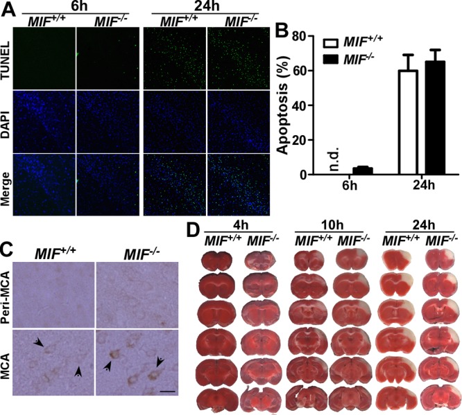
Figure 5.

MIF reduced cell death and infarct development during stroke. A) Wild-type (Mif+/+) and MIF-knockout (Mif −/−) mice were subjected to 2 h tMCAl, followed by 4 or 22 h reperfusion, and the brain slices were analyzed for cell apoptosis by TUNEL assay (green channel). Nuclei were stained with DAPI (blue channel). Representative images were taken from the same location in the MCA territory. B) Average counts of the nuclei and TUNEL-positive cells from 5 selected views were quantified by ImageJ; bars represent percentage of TUNEL signal. Values represent means ± sd, n = 3. P > 0.05; Student's t test. C) Cleaved caspase-3 was determined histologically in wild-type and MIF-knockout mice 4 h after 2 h tMCAl. Sample images were taken from the cortical peri-MCA region and MCA territory. Activation of caspase-3 is indicated by positive staining of the cleaved form of caspase-3. Arrows indicate caspase-3-positive cells. Scale bar = 20 μm. D) Mif+/+ and Mif−/− mice were subjected to 2 h tMCAl on the right hemisphere, followed by 2, 8 or 22 h reperfusion. The brain was freshly cut into 1 mm slices and subjected to TTC staining.
