
Figure 2. Mutation of Pparγ at K154/155 reduces SIRT1 binding.
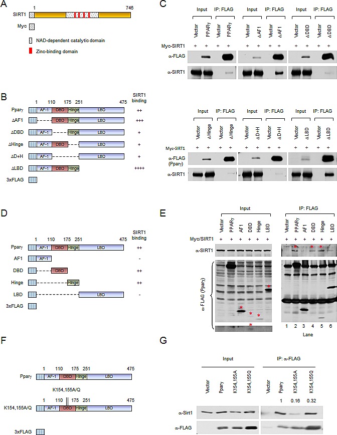
(A) Schematic diagram of Myc-tagged SIRT1 wild-type. (B) Schematic diagrams of 3xFLAG-tagged Pparγ1 full length and internal deletion mutants. (C) HEK 293T cells were transfected with the indicated plasmids. Immuno-precipitations with anti-FLAG antibody were conducted, and Western blot was performed by indicated antibodies. The abundance of SIRT1, PPARγ internal deletions in input are shown. (D) Schematic diagrams of 3xFLAG-tagged Pparγ1 full length and individual domains. (E) HEK 293T cells were transfected with the indicated plasmids. Immuno-precipitations with anti-FLAG antibody were conducted, and Western blot was performed by indicated antibody. The bands for 3x FLAG-tagged Pparγ full length and individual domains are indicated by an asterisk. (F) Schematic diagrams of 3xFLAG-tagged Pparγ1, Myc-tagged SIRT1 wild-type and catalytic point mutation. (G) HEK 293T cells were transfected with the indicated plasmids. Immuno-precipitations with anti-FLAG antibody were conducted, and Western blot was performed by indicated antibody. All experiments were performed at least 3 times, representative figures are shown.