
Figure 4. Src/FAK and associated proteins are selectively activated in the mesenchymal cells, and are suppressed by dasatinib.
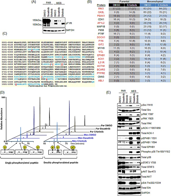
(A) Immunoblot demonstrating suppression of a phospho-pY band at 140kDa following treatment with dasatinib (30nM) but not erlotinib (50nM) in the mesenchymal HCC827 cells at 24h. (B) Table of peptide spectral matches (PSMs) total (unique) for phosphotyrosine peptides following erlotinib (50nM) treatment for 24h, demonstrating the most significant changes in phosphorylation in the HCC827 mesenchymal cells. Highlighted in red are those kinases associated with Src/FAK signaling. (C) Mass spectrometry-derived peptide sequence coverage of FAK after phosphotyrosine containing PSMs mapping to FAK. Identified peptide sequences are noted in blue and confidently localized phosphorylation sites are denoted in red. (D) Extracted ion chromatograms of various singly and doubly phosphotyrosinated peptides from sites Y570/Y576/Y577 of FAK showing relative intensities of each species across experimental conditions. (E) Immunoblot demonstrating suppression of phospho-FAK, pACK-1, pEPHB1, phospho-p38 and phospho-STAT3 following treatment with dasatinib (30nM) and not erlotinib (50nM) in the mesenchymal HCC827 cells at 24h.