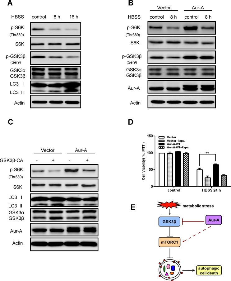
Figure 5. Aur-A activated mTOR signaling by inhibiting GSK3β under metabolic stress.
A-B, mTOR activity (indicated by p70S6K phospho-Thr389 level) and GSK3β activity (indicated by phospho-Ser9 level) were assessed by Western blot after HBSS starvation in control and Aur-A overexpressed SKBR-3 cells. C, GSK3β activity was reconstituated by transfection of V5-GSK3β-CA plasmid in control and Aur-A overexpressed SKBR-3 cells. After HBSS starvation, mTOR activity (indicated by p70S6K phospho-Thr389 level) and LC3 conversion (LC3 II/total) were assessed by Western blot. D, Cells were pretreated with rapamycin 12 hours before subjected to HBSS treatment. After starvation, cell viabilities were measured by MTT assay. Data were mean ± S.D. of three independent experiments done in parallel (**p<0.01). E, A working model to elucidate signaling pathway that mediated action of Aur-A under metabolic stress.

