
Figure 6. Increased P450R expression renders B16-MDSCs susceptible to Paclitaxel.
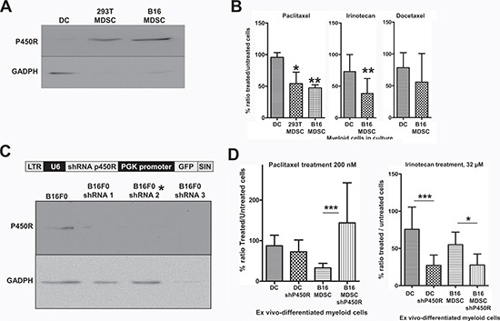
(A) Detection of P450R by immunoblot in DCs, 293T-MDSCs and B16-MDSCs as indicated. GADPH expression was used as a loading reference. P450R was very highly expressed in MDSCs compared to DCs (of note, GADPH is expressed at the same levels according to quantitative mass spectrometry). (B) Bar graphs representing the ratio (as a percentage) of drug-treated versus untreated DCs, 293T-MDSCs and B16-MDSCs after an overnight treatment with the indicated chemotherapy drugs, at known cytotoxic concentrations for cancer cells. Error bars correspond to standard deviations. (C) The lentivector platform used to deliver different P450R-silencing shRNAs is shown above. Below, detection of P450R or GADPH by immunoblot in B16F0 cells transduced with three P450R-silencing lentivectors. The P450R-shRNA2 lentivector was used to modify myeloid cells, indicated with an asterisk. (D) Bar graph on the left represents the ratio of paclitaxel-treated versus untreated DCs and B16-MDSCs, unmodified or in which P450R was silenced, as indicated. The bar graph of the right shows the same type of experiments but using Irinotecan to treat cell cultures. LTR, long-terminal repeat; U6, U6 promoter; shRNA, short-hairpin RNA coding sequence; PGK, phosphoglycerolate promoter; SIN, self-inactivating LTR. *, **, ***, indicate significant, very significant and highly significant differences, respectively.