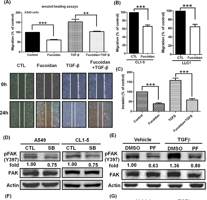
Figure 7. Fucoidan inhibits the phosphorylation of FAK and suppresses the TGFβ-dependent migratory and invasive properties of lung cancer cells.
(A) Wound closure assays for the effect of fucoidan on A549 cells. Images were captured at 0 and 24 h in serum-free medium with TGFβ (10 ng/ml) or without (control) in the presence of vehicle (PBS) or fucoidan (200 μg/ml). Total magnification: × 40. (B) To assess the migration potential, the highly metastatic CL1-5 and LLC1 cells were treated with fucoidan (200 μg/ml) for 24 h and then subjected to the Transwell® assay. (C) A549 cell invasion was assayed in the presence of fucoidan alone (200 μg/ml) or fucoidan in combination with TGFβ (10 ng/ml). A549 cells (5×10⁵ cells/well) were seeded into the upper chamber with DMEM, with the bottom chamber coated with Matrigel® and the lower chamber containing DMEM with 10% fetal bovine serum (FBS). The cell migration and invasion assays were performed for 24 h in a humidified incubator at 37°C with 5% CO2. Migratory and invading cells were counted under the microscope after fixation and staining with crystal violet. The data are expressed as the percentage of migration or invasion through the membrane or Matrigel® matrix. The numbers of cells that migrated were observed and counted using an Olympus IX70 Inverted Microscope and SPOT Advanced Digital Imaging. One representative experiment of three independent experiments is shown. (D) The TGFR inhibitor SB431542 (SB) down-regulates the phosphorylation of FAK. A549 and CL1-5 cells were cultured in serum-free media for 24 h, followed by treatment with vehicle (-) or SB (20 μM) for 3 h and western blot analysis. (E) The FAK inhibitor PF573228 (PF, 2 μM) inhibits the TGFβ-induced phosphorylation of FAK. (F) PF573228 abolishes the TGFβ (10 ng/ml)-induced migration for 24 h in A549 cells. (G) Fucoidan suppresses the TGFβ-induced phosphorylation of FAK. A549 cells were cultured in serum-free media for 24 h and then treated with vehicle (-) or PF (2 μM) in (E) or with fucoidan (Fu, 200 μg/ml) in (G) for 30 min. The cells were then incubated with TGFβ (10 ng/ml) for 3 h and then subjected to western blot analysis. The proteins of whole cell lysates were immunoblotted with anti-pFAK (pY-397). FAK and actin served as internal controls for loading. One of three individual experiments is presented. Significant differences are shown (*P<0.05, **P<0.01 and ***P<0.005, compared with the control group).

