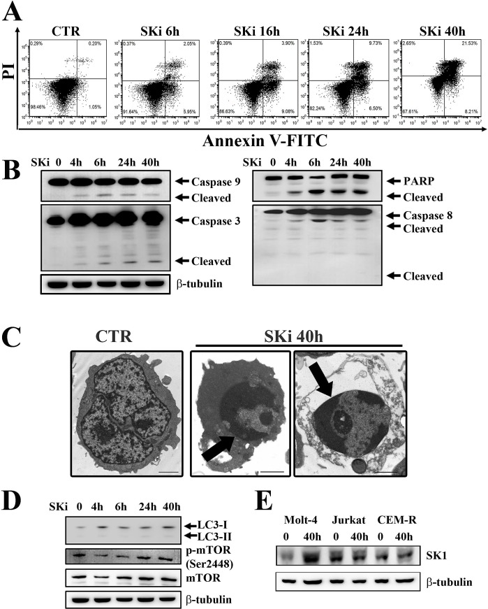
Figure 2. SKi triggers apoptosis in Molt-4 cell line.
(A) Flow cytometric analysis of Annexin-V-FITC/PI stained Molt-4 cells treated with SKi (6.9 μM) for 6, 16, 24 and 40 h documented a time-dependent increase in apoptotic cells with respect to untreated cells. (B) After SKi treatment, cells were collected, lysed and analyzed by western blotting for cleaved caspase-9, -3, and -8. (C) Molt-4 cells were treated with SKi (6.9 μM) for the indicated time, then processed for TEM analysis that documented apoptosis induction. Arrows point to condensed apoptotic chromatin. Scale bar 2 μm. (D) Western blot analysis for the autophagic marker LC3 and p-mTOR (Ser2448) in cells treated with SKi for the indicated times. (E) Western blot analysis for SK1 expression in T-ALL cell lines upon SKi treatment for 40 h. In (B) (D) and (E) 50 μg of protein was loaded for each lane. β-tubulin was used as a loading control.

