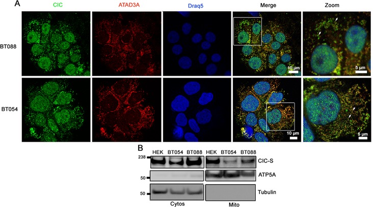
Figure 4. CIC-S localizes to mitochondria in 1p19q co-deleted oligodendroglioma cell lines.
A. Subcellular localization of CIC in ODG cell lines BT088 and BT054 was detected using immunofluorescence. CIC was detected using an anti-CIC antibody and mitochondria were stained using an anti-ATAD3A antibody. Cells were visualized using confocal fluorescence microscopy. B. Cytosolic (Cytos) and mitochondrial (Mito) fractions of BT054 and BT088 were isolated and CIC-S was detected using western blots. Control proteins ATP5A (Mito) and Tubulin (Cytos) were also detected.

