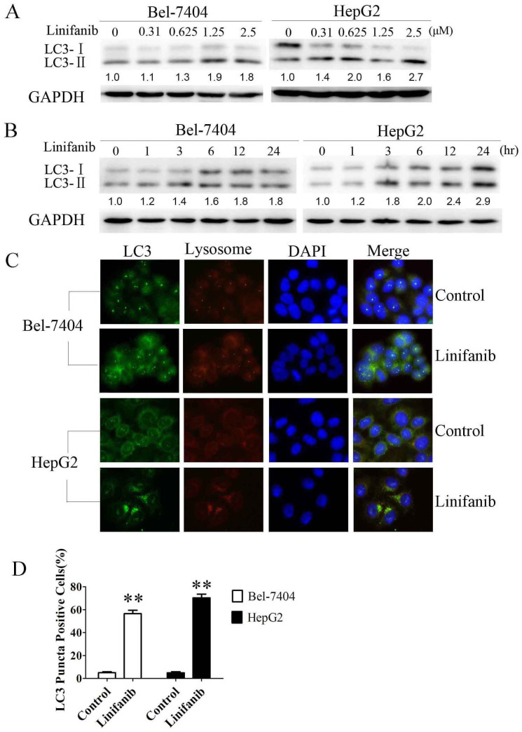
Figure 1. Effect of linifanib on LC3 processing.
(A) & (B). Cells were incubated with the indicated concentration of linifanib for 24 h or incubated with 2.5 μM linifanib for the indicated intervals, and the transition of LC3-I to LC3-II was analyzed by Western blotting. Relative quantity of LC3-II was calculated by ImageJ densitometric analysis and normalized by GAPDH. (C). Cells were treated with DMSO or 2.5 μM linifanib for 24 h before they were labeled with fluorescence and imaged by fluorescence microscope. Green: FITC-labeled LC3; Red: lyso-tracker-labeled lysosome; Blue: DAPI-labeled nucleus. (D). Percentage of green or yellow puncta-positive cells was quantified and analyzed using a threshold of >5 dots/cell. The data were represented as the mean ± SD. **P < 0.01.

