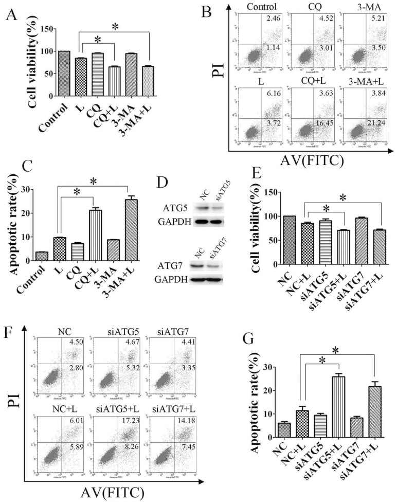
Figure 5. Inhibition of autophagy promotes linifanib-induced apoptosis in vitro.
(A). Bel-7404 cells were treated with 2.5 μM linifanib (L) or DMSO in the presence or absence of CQ (5 μM) or 3-MA (5 mM) for 24 h. Cell viability was measured by MTS assay. (B) & (C). Cells were treated with linifanib alone or in combination of CQ or 3-MA before staining with annexin V (AV) and propidium iodide (PI) followed by flow cytometry. (D to G), Bel-7404 cells were transfected with siRNA against ATG5 or ATG7 for 24 h, then treated with linifanib for 24 h. Cell viability was measured by MTS assay (E), and apoptotic rates were determined by staining with annexin V and propidium iodide followed by flow cytometry (F & G). Data were expressed as the mean ± SD. * P<0.05.

