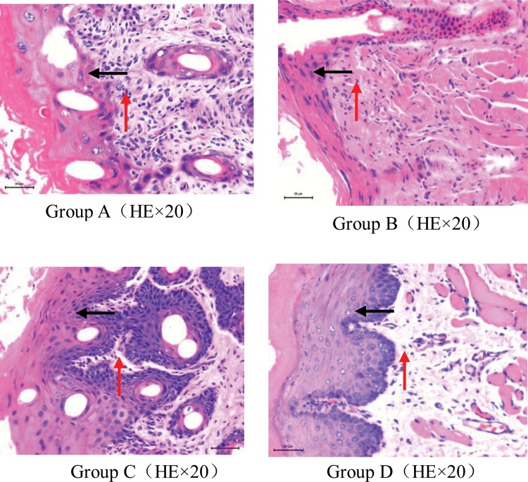
Fig. 3.
Representative and typical microscopic changes in the buccal mucosa of guinea pigs in each group on Day 8 after irradiation. Group A. The epithelial layer was destroyed, and the structure was not clear (black arrow). Epithelium hyperplasia (black arrow), interstitial proliferation, and variable inflammatory cell infiltration were observed (red arrow). Sometimes, ulcer formation was observed. Group B. Destruction of the epithelium and moderate inflammatory cell infiltration in the stroma were observed, however, the degree of injury of the tissues was less than in Group A. Group C. The epithelial structure was heavily damaged. Ulcer formation, significant dysplasia of the epithelial and interstitial tissues, and substantial inflammatory cell infiltration were observed. Group D. Intact, healthy mucosa.

