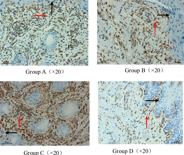
Fig. 4.
The intensity of the expression of ICAM-1 of the buccal mucosa and stroma of guinea pigs on Day 8 after irradiation. Weak constitutive ICAM-1 expression (staining intensity 1) in the epithelium (black arrow) and stroma (red arrow) of a sham-irradiated control buccal mucosa was shown in Group D. Group C. Increased ICAM-1 expression (staining intensity 3) in the epithelium and stroma on Day 8 after irradiation. The increase in ICAM-1 expression was accompanied by substantial leukocyte infiltration, epithelium hyperplasia and stroma proliferation. The staining intensity of ICAM-1 expression in the vast majority of specimens in Group A and B was at a level of 2, and a few were stained as 3 or 1.

