
Fig. 1.
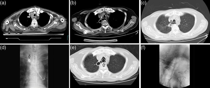
A patient with esophageal cancer who received radiotherapy after exploratory thoracotomy, (a) CT image when started the RT, the arrow head showed an extracapsular LN invading the esophagus, (b) CT image (mediastinal window) of the esophageal perforation (the arrow head indicates the actual communication of an air-filled esophagus with an adjacent mediastinal or paramediastinal air–fluid collection), (c) the perforation on lung window, (d) the leakage of iodine on iodine examination, (e) CT image after the placement of a self-expandable metallic stent (the arrow head indicates the stent), and (f) an X-ray after the placement of a stent (the arrow head indicates the stent).