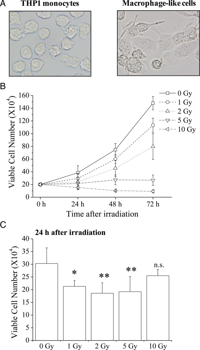
Fig. 1.

Cell morphology and viable cell numbers of X-irradiated THP1 monocytes and macrophage-like cells. (A) Morphology of THP1 monocytes (left) and macrophage-like cells (right). (B) THP1 monocytes were exposed to X-irradiation and were cultured for 24–72 h. Cells were harvested and viable cell numbers were estimated using the trypan blue dye exclusion method. (C) Macrophage-like cells were exposed to X-irradiation and were cultured for 24 h. Cells were harvested and viable cell numbers were estimated using the trypan blue dye exclusion method. Data are presented as the mean ± SD of four independent experiments; single and double asterisks indicate P < 0.05 and P < 0.01 compared with non-irradiated control, respectively; n.s. = not significant.
