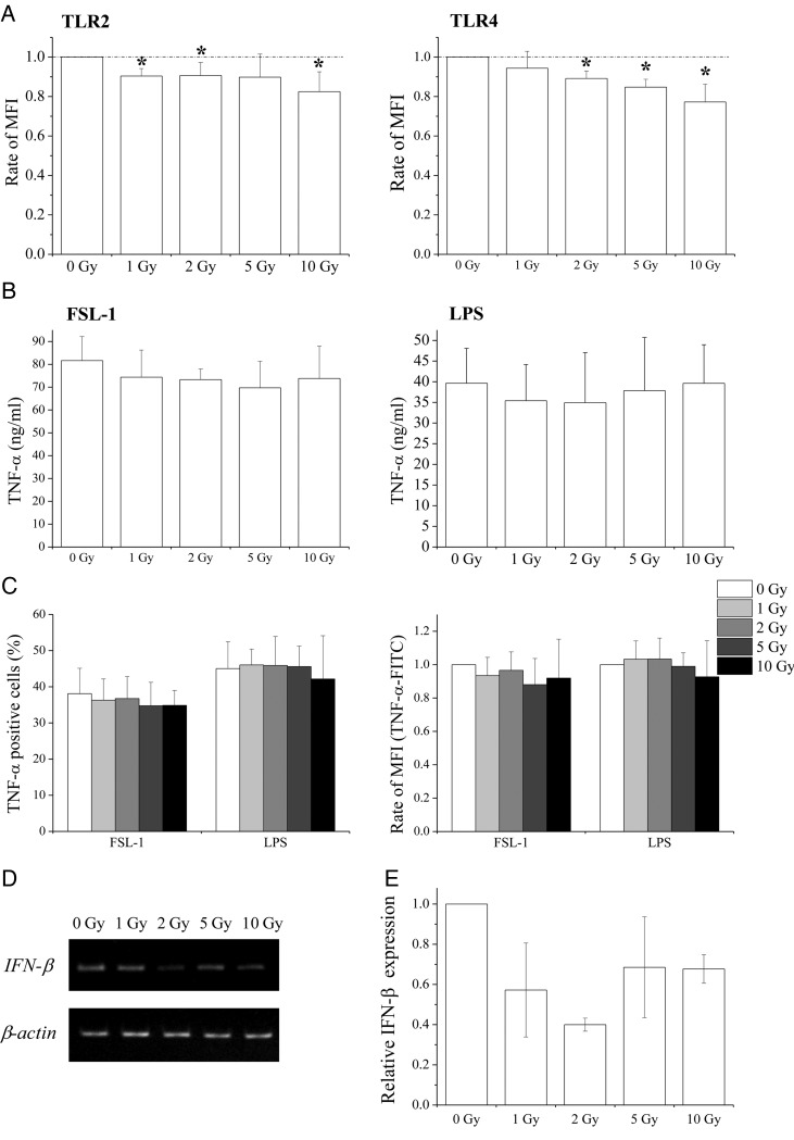
Fig. 3.
Effects of X-irradiation on TLR2 and TLR4 expression and response to ligands in macrophage-like cells. (A) Non- or X-irradiated macrophage-like cells were cultured for 24 h and the expression of TLR2 and TLR4 was analyzed using flow cytometry. Data are presented as the mean ± SD of four independent experiments; single asterisk indicates P < 0.05 compared with non-irradiated control. (B) Non- or X-irradiated macrophage-like cells were cultured for 24 h, and FSL-1 or LPS were added to culture supernatants. After an additional 24 h of culture, culture supernatants were harvested and TNF-α concentrations were determined using ELISA. Data are presented as the mean ± SD of four independent experiments. (C) Non- or X-irradiated macrophage-like cells were cultured for 24 h, were then stimulated with FSL-1 or LPS for 8 h, and the expression of intracellular TNF-α was analyzed. Data are presented as the mean ± SD of four independent experiments. (D, E) Non- or X-irradiated macrophage-like cells were cultured for 24 h and LPS was added to culture supernatants. After an additional 24 h of culture, RNA was extracted from cells and the expression of IFN-β was determined using RT-PCR (D) and real-time quantitative RT-PCR (E) analyses. Data from real-time quantitative RT-PCR experiments are presented as the mean ± SD of three independent experiments.

