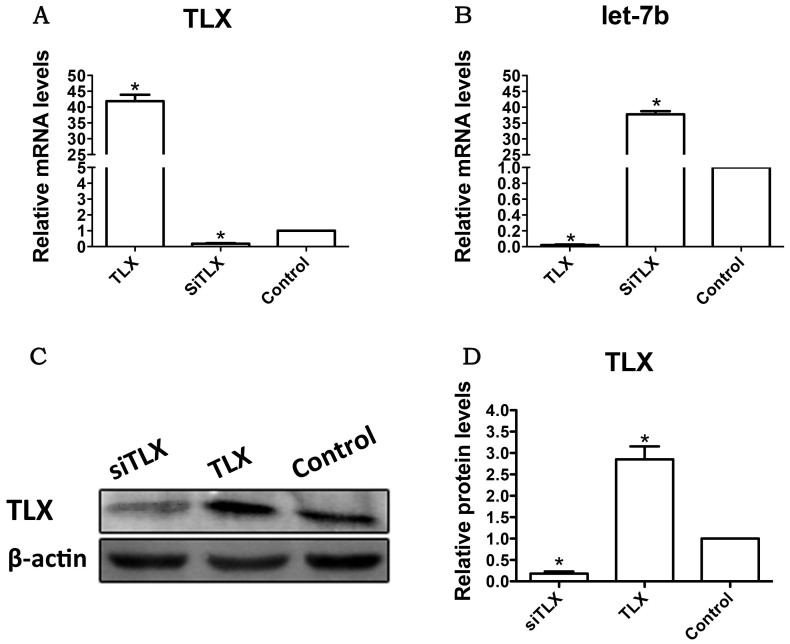
Figure 5. Effects of the overexpression or inhibition of TLX on the expression of let-7b in the RPC cultures.
(A): The qPCR results revealed that the expression level of TLX decreased sharply with siTLX treatment but increased approximately 40-fold in the TLX clone-treated RPC cultures compared with the control. (B–D): The expression level of let-7b was markedly increased in siTLX-treated RPC cultures and decreased in the TLX clone-treated cells, a result that was consistent with that from Western blot analysis. Error bars indicate the standard deviation of the mean; *p < 0.05 by Student's t-test. Full-length blots/gels are presented in Supplementary Figure 8.

