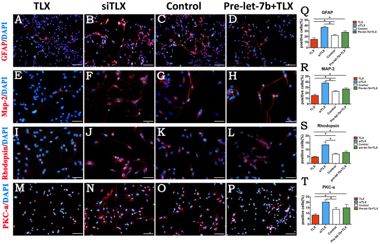
Figure 7. TLX inhibits the differentiation of RPCs.
(A–T): RPCs transfected with TLX and immunostaining with antibodies against GFAP, MAP-2, rhodopsin, and PKC-α, the proportions of GFAP-, MAP-2-, rhodopsin-and PKC-α- immunoreactive cells were detected to be higher in the siTLX-treated cultures than in the control cells, but their positive percentage decreased by TLX clone treatment. Treatment with let-7b rescued the reduction, which was caused by the overexpression of TLX. Error bars indicate the standard deviation of the mean; *p < 0.05 by Student's t-test. Scale bars: 200 μm (A–B), 50 μm (E–L), 100 μm (M–P).

