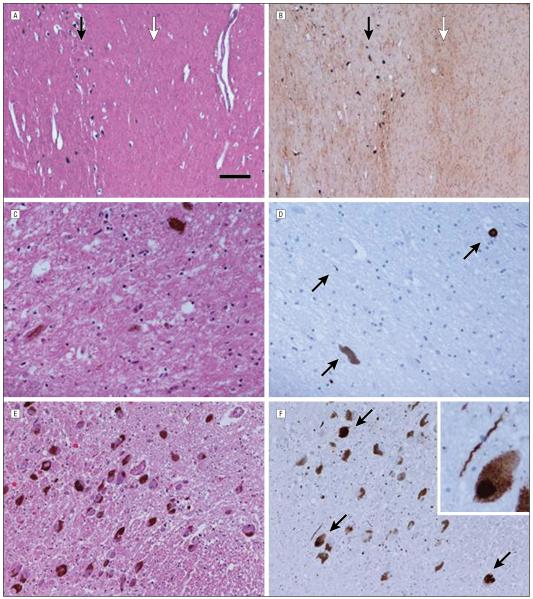
Figure 1. Histologic findings in the parkin cases.
Case 2 substantia nigra illustrates the more severe neuronal loss in the ventral tier (white arrow) compared with the dorsal tier (black arrow) (A), accompanied by severe gliosis (white arrow) in the ventral tier compared with the dorsal tier (black arrow) (B). Case 5 showed severe neuronal loss in the ventrolateral substantia nigra (C) and only sparse α-synuclein pathology (arrows) (D), with mild cell dropout in the locus coeruleus (E) accompanied by moderate numbers of Lewy bodies (arrows) and Lewy neurites (F). The scale bar represents 260 μm in parts A and B, 50 μm in parts C and D, 100 μm in parts E and F, and 25 μm in the inset in part F. Hematoxylin-eosin staining in parts A, C, and E; glial fibrillary acidic protein in part B; and α-synuclein in parts D and F.

