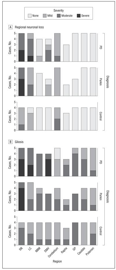
Figure 2. Regional neuronal loss (A) and gliosis (B) in parkin, Parkinson disease (PD), and control cases.
A, The severity of neuronal loss in various brain regions in the 5 parkin, 5 PD, and 4 control cases (not all structures were available for examination in all cases [eg, the raphe was only available for examination in 3 parkin, 2 PD, and 3 control cases]). B, The severity of gliosis in various brain regions in the parkin, PD, and control cases. GP indicates globus pallidus; DMV, dorsal motor nucleus of the vagus; LC, locus coeruleus; NBM, nucleus basalis of Meynert; SN, substantia nigra.

