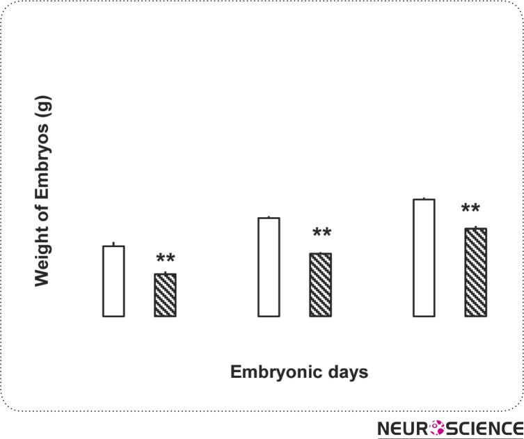
Figure 3.
It is suggested that Micrograph of section of spinal cord of 14-days-old embryo in control group(A). W.M: White matter, G.M: Gray matter, Vent: Ventricular layer, E.C: section of spinal cord of 12-days-old embryo in experimental group(B). W.M: White matter, G.M: Gray matter, Vent: Ventricular layer, E.C: Ependimal channel (×100).

