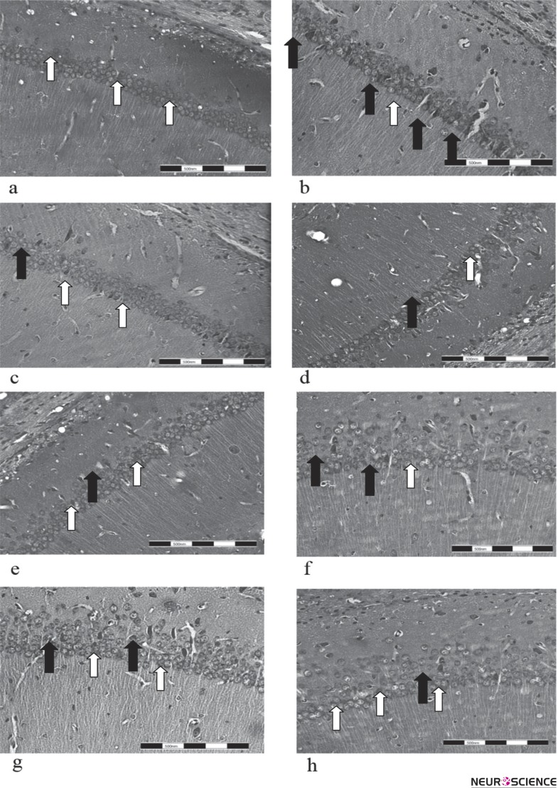
Figure 2.
Neuronal death in CA1 region of hippocampus
Cresyl violet staining of brain sections from experimental groups was performed at the end of treatment used to evaluate the neuronal density and structure. The Animals treated with CPA (f) or vitamin C (g) had more cell density which is representative of less dead neurons compared to the ischemia group (b). Co-administration of both CPA and vitamin C reduces cell death (h) compared to ischemia groups and groups received CPA (f) or vitamin C (g). Administration of A1 receptor antagonist (DPCPX) intensified the cell death among ischemic neurons and reduced cell density (d). White and black arrows are representative of normal and dead cells, respectively. Experimental groups including Intact, Ischemia, DMSO, DPCPX, DPCPX/AA, CPA, AA (vitamin C) and CPA/AA are shown a, b, c, d, e, f, g, h digital images prepared at 40 x magnifications, respectively. Scale bars: 200 µm.

