Figure 1.
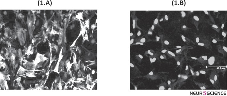
The purity of astrocyte cultures was determined using indirect immunocytochemical staining with anti-GFAP antibody. (1.A) Confocal Microscopy analysis of primary astroglial cells stained with rabbit anti-GFAP antibody (green). The cell nuclei stained with DAPI (blue). (1.B) Negative control stained with secondary antibody and DAPI. Immunocytochemistry for determination of GFAP, were done for each astrocyte cultures.

