
Figure 4.
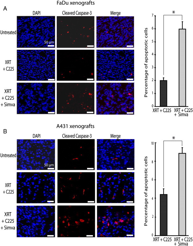
Simvastatin was associated with apoptosis in xenografts. Mice were treated with XRT and C225 or with XRT, C225, and simvastatin as described in Figure 1. Tumors were extirpated on the fourth day, 24 hours after the last treatment. Representative pictures showing cleaved caspase-3 (red fluorescence) and cell nucleus (blue fluorescence) are shown for FaDu- (A) and A431-derived (B) tumors. Columns show the mean values and bars show the SEM of apoptotic cells relative to the total number of cells. Six randomly selected field microscopic images were analyzed per slice. On average, 200 cells were counted per field. Data were obtained from two independent experiments from each treatment scheme. *P < .05 compared to XRT plus C225.