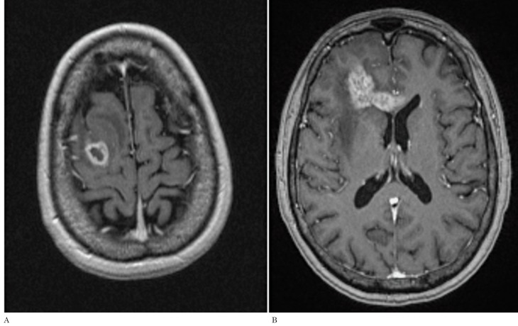
Figure 3.
A) Preoperative axial postcontrast T1-weighted image of an enhancing GB which is in contact with the subcortical fronto-parietal region (Group II). B) Post-treatment T1-weighted image of the same patient demonstrating a ‘distant’ type of recurrence located in the genu of corpus callosum and right frontal lobe that represents the least common type of recurrence found in our analysis

