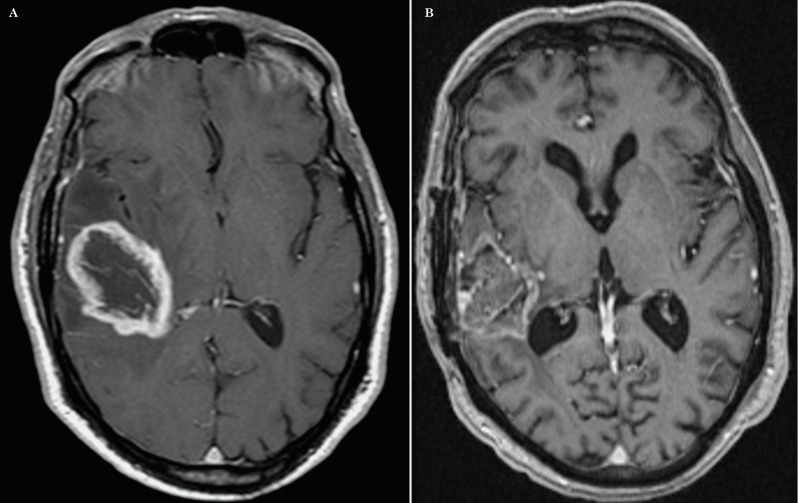
Figure 4.
A) Axial postcontrast T1-weighted image of an enhacing GB prior to any treatment which is in contact with the subventricular zone of the right lateral ventricle atrium and also in contact to the temporal subcortical region (Group III). B). The post-treatment T1-weighted image illustrates the enhancing tumor within the original surgical bed thus being classified as a ‘local’ recurrence type. This was the most common recurrence type for this group.

