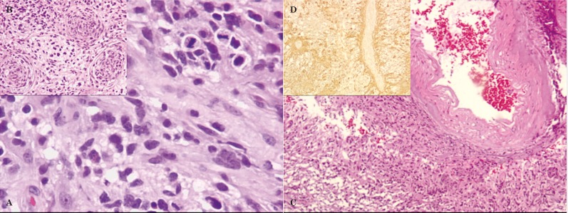
Figure 5.
Histopathology, after partial resection of right anterior temporal lobe (18th day) - glioblastoma multiforme (A-C: HE; D: GFAP). A) Highly cellular neoplasia with hyperchromatic pleomorphic nuclei and mitosis. B) Three tumoral vessels with prominent vascular wall proliferation. C) Tumor involving and infiltrating the leptomeningeal vessels. D) Tumor infiltrating an intracortical vessel.

