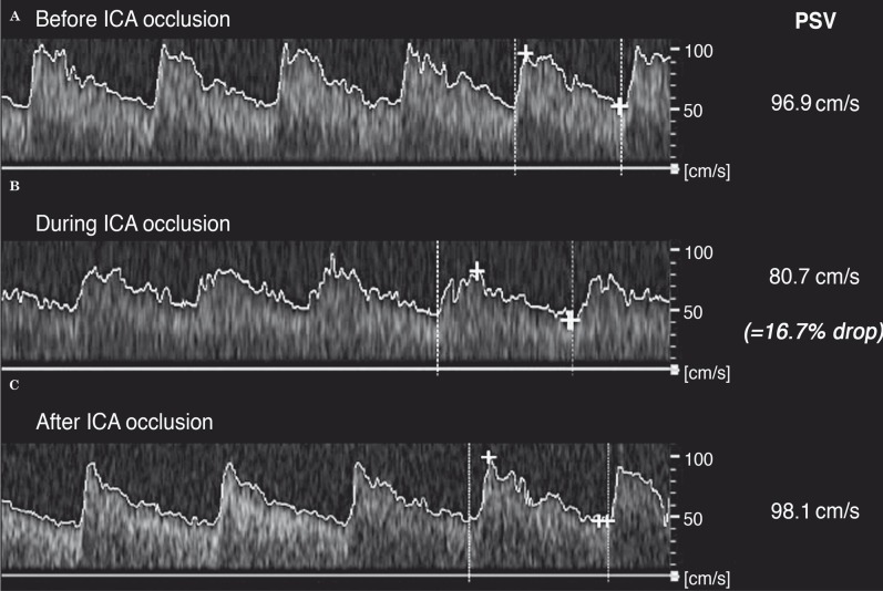
Figure 3.
Angle corrected left middle cerebral artery flow variation registered by transcranial Doppler during the procedure. A) Before left ICA occlusion. B) During ICA occlusion. C) After balloon deflation. Note the documented 16.7% decrease of the peak systolic velocity of the middle cerebral artery (PSV), inferior to the 30% threshold.

