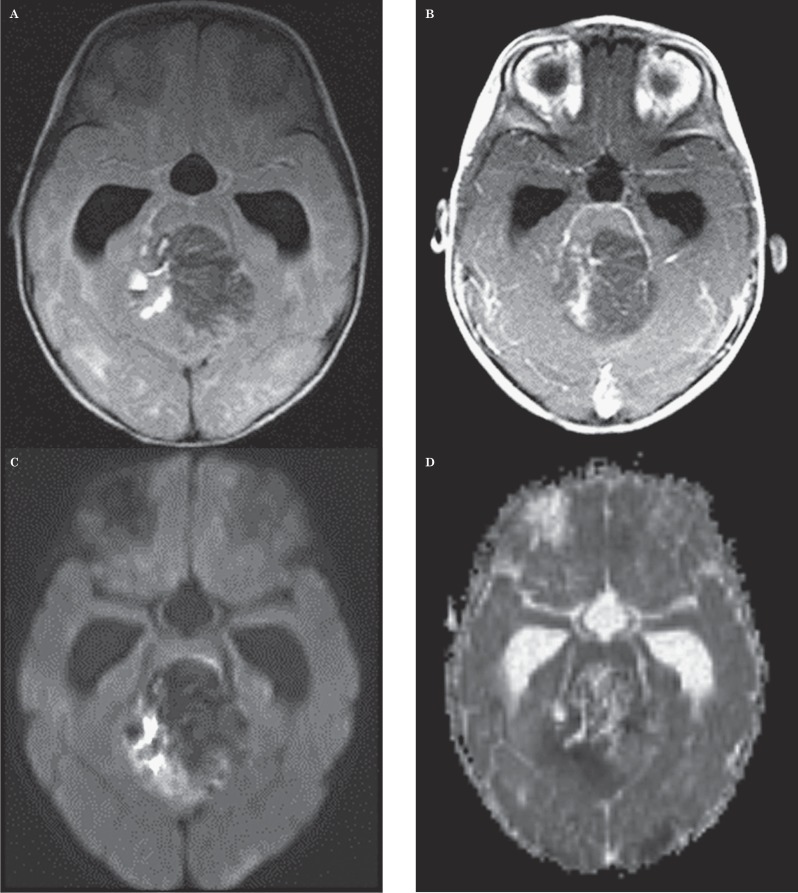
Figure 2.
Seventeen-year-old girl with an anaplastic astrocytoma. This case represents the single MR imaging study in which both neuroradiologists provided the correct diagnosis and in which ADC values indicated an incorrect diagnosis. However, the location and appearance are atypical for medulloblastoma. A) Axial FLAIR image shows a complex mass involving the pons, left middle cerebellar peduncle and left cerebellar hemisphere. This case is the sole one in which, of the 6 cases that would have been incorrectly diagnosed by the ADC threshold, the clinical impression of both radiologists would have been correct. This outcome suggests that weighting the ADC threshold more heavily in the interpretation of imaging of PFT would rarely result in a less accurate diagnosis than clinical impression alone. B) Axial contrast-enhanced T1-weighted image shows a contrast-enhancing mass containing multiple cystic regions. C) Axial DWI image shows that the portion of the tumor within the pons has bright signal, possibly indicating restricted diffusion. D) Axial ADC map at the same level as C shows the pontine lesion has a dark signal suggestive of low ADC values. Mean ADC value measured by one observer was 0.615 mm2/s and that by the other observer was 0.624 mm2/s, suggesting the diagnosis of medulloblastoma. However, the imaging features shown in A and B would be atypical for medulloblastoma based on lesion location (i.e., pontine involvement) and the presence of a large cyst.

