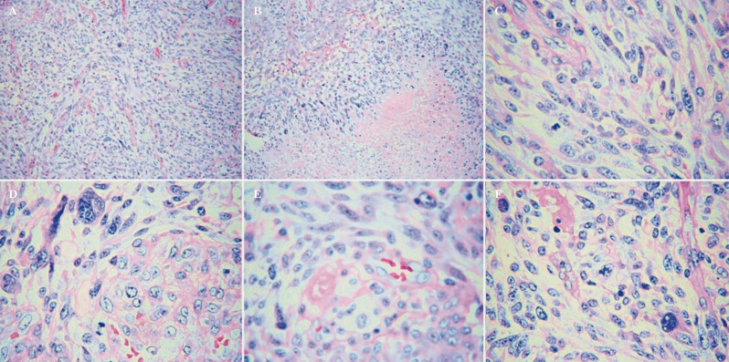
Figure 2.
Histopathological studies of all tumors identified a mixed CNS neoplasm with large areas of spindle cell differentiation (A, 100×) and extensive areas of necrosis (B, 100×). The neoplastic cells showed a highly pleomorphic morphology (C, 100×), some were multinucleated (D, 400×). Mitotic figures and microvascular proliferation were frequently seen in all cases (E, F, 400×).

