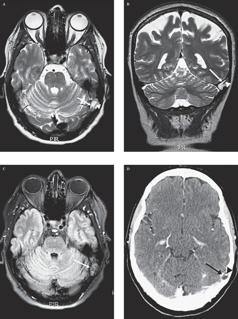
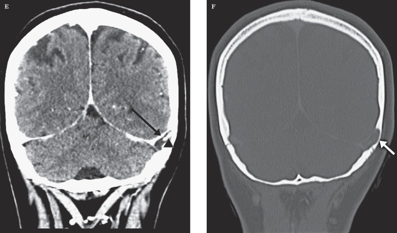
Figure 1.
Patient 1. Giant arachnoid granulation in the left transverse sinus. Transverse (A) and coronal (B) T2-weighted images. An ovalar hyperintense structure (arrow) can be seen in the left transverse sinus at the transition to the sigmoid sinus. Intralesional flow void (arrowhead). C) Transverse FLAIR. There is attenuation of the content of this “lesion” (arrow) that remains hyperintense. Transverse (D) and coronal (E) enhanced CT. The “lesion” (arrow) is hypodense with a central enhancing vascular structure (arrowhead). E) Coronal CT with bone windows. Note the scalloping of the overlying bone (arrow).


