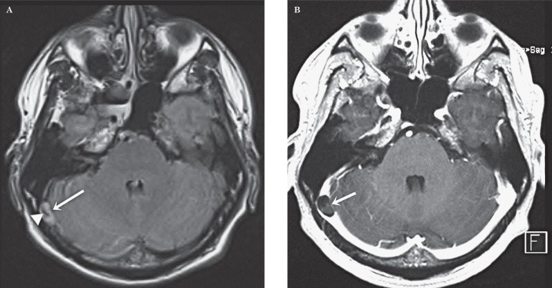
Figure 2.
Patient 2. Giant arachnoid granulation in the right transverse sinus. A) Transverse FLAIR. Note the hyperintense “lesion” (arrow) in the left transverse sinus at the transition to the sigmoid sinus. Central flow void pointing to a vascular structure (arrowhead). B) Transverse gadolinium-enhanced image. The hypointense ovalar lesion (arrow) in the transverse sinus is confirmed. The vascular structure less well visible due to volume-averaging.

