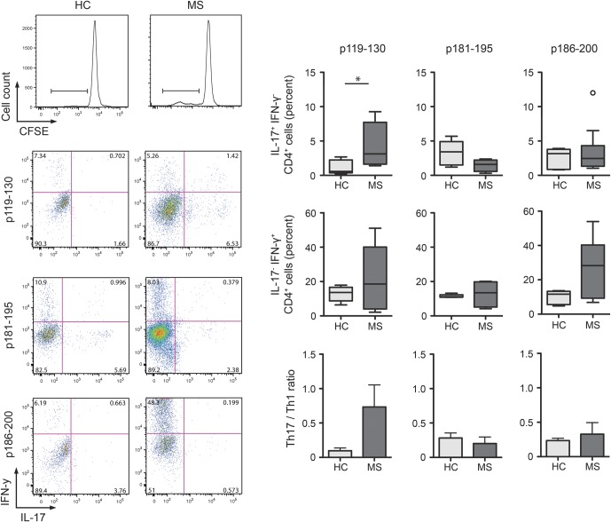
Figure 2. T cells from patients with multiple sclerosis (MS) exhibit a proinflammatory bias.
CD4+CFSElow proliferating T cells (cell division index >2) were analyzed for interleukin (IL)-17 and interferon (IFN)-γ production by intracellular staining after stimulation with phorbol 12-myristate 13-acetate/ionomycin for 5 hours. Frequencies of IL17+IFN-γ−, IL17+IFN-γ+, and IL17−IFN-γ+ were examined among proliferating p119-130–specific CD4+ T cells, p181-195–specific CD4+ T cells, and p186-200–specific CD4+ T cells. Frequencies of IL-17 and IFN-γ single positive T cells were used to calculate Th17/Th1 ratio. Box and whisker plots include the median, distribution, and range. *p < 0.05, Mann–Whitney U test. CFSE = carboxylfluorescein diacetate succinimidyl ester; HC = healthy controls.

