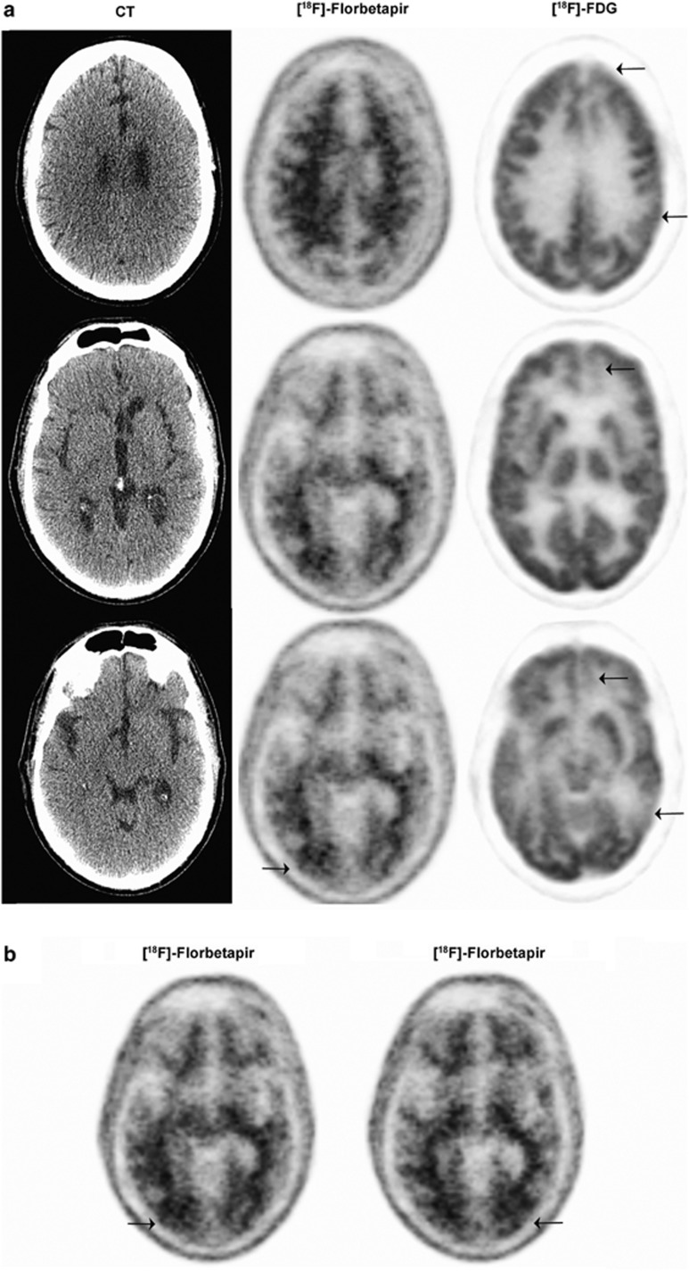
Figure 4.
Imaging from a 59-year-old, physician with a sports-related injury (case 2). [18F]-Florbetapir PET imaging findings were negative for amyloid accumulation except for focal [18F]-Florbetapir retention at the site of impact in the occipital region (arrows). (a) CT (left panel) [18F]-Florbetapir PET (middle panel) and FDG PET (right panel) at various depths of the brain. (b) [18F]-Florbetapir PET indicating amyloid accumulation. CT, computed tomography; FDG, [18F]-fluorodeoxyglucose; PET, positron emission tomography.

