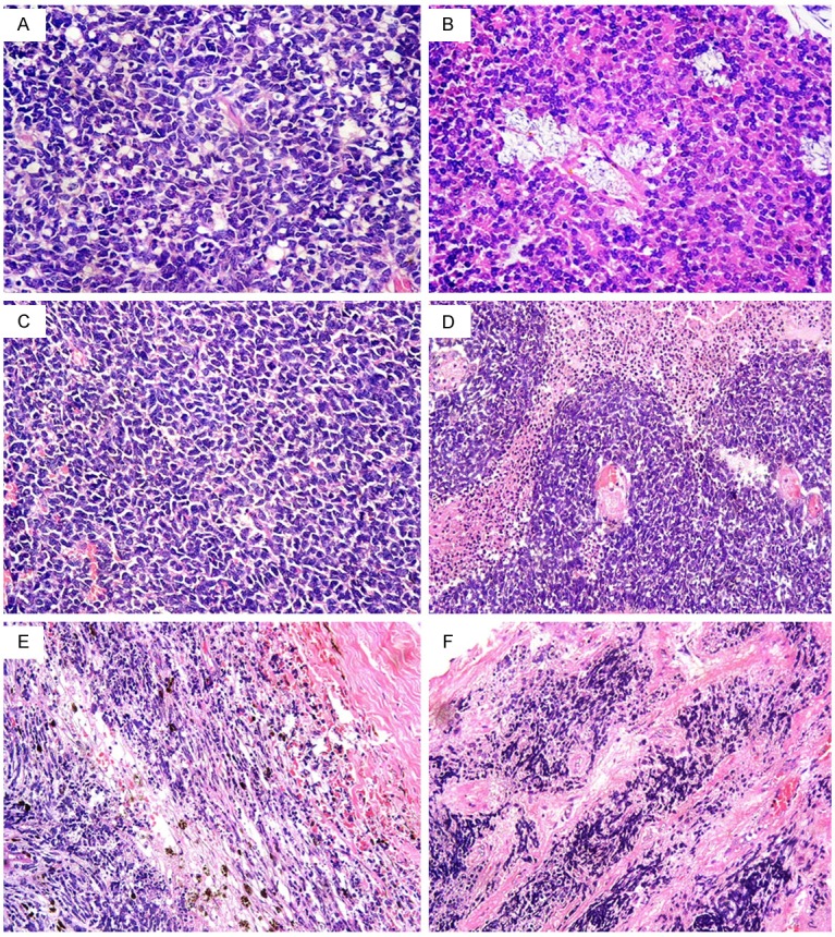
Figure 1.

Retinoblastoma: (A) Well differentiated tumour with foci of Flexner-Wintersteiner rosettes, (B) Well differentiated tumour with foci of Flexner-Wintersteiner rosettes and fleurettes, (C) Poorly differentiated tumour, (D) Area of retinoblastoma illustrating the sleeve pattern of growth around central blood vessels. The three cuffs of viable neoplastic cells are well demarcated from the surrounding necrotic cells, (E) Massive invasion of the choroid, (F) Post laminar optic nerve invasion [hematoxylin-eosin, original magnification, (A-C) × 400; (D-F) × 200].
