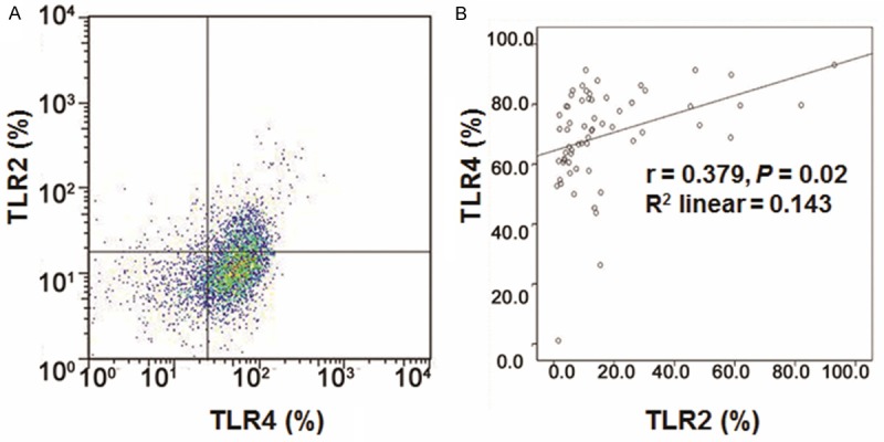
Figure 2.

Expression of TLR2 and TLR4 in DCs in HBV patients. A. Flow cytometry was applied to detect the expressions of TLR2 and TLR4 in DCs. Cells in Q1, Q4 were considered as negative for TLR2 and TLR4 respectively, while Q2 and Q3 were considered as positive for TLR2 and TLR4, respectively. B. TLR2 and TLR4 were positively correlated (r = 0.379, P = 0.02).
