
Figure 1.
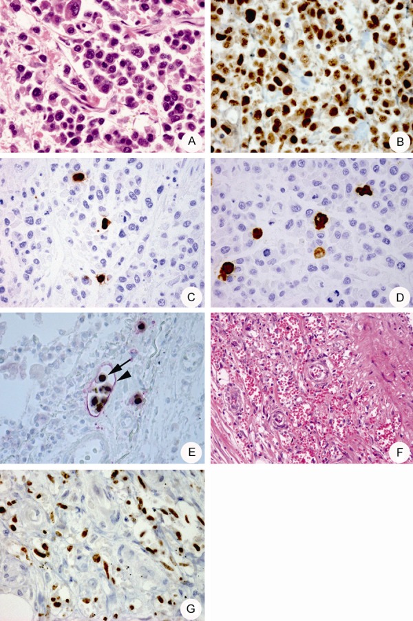
(A) Microscopic morphology of a ureter tumor, following hematoxylin and eosin (HE) staining. 400× magnification. (B) HHV-8 LANA-1 detection in tumor cells with punctate staining. 400× magnification. (C, D) A small number of ORF59 (C) and vIL-6 (D) positive tumor cells are observed. 400× magnification. (E) Histology of a lymphoma cell embolism in the pleural lymphatic vessels with double immunohistostaining using antibodies against HHV-8 LANA-1 (arrow) or D2-40 (arrowhead). 400× magnification. (F) Histology of the Kaposi’s sarcoma (KS) in the ureter (HE stain). 100× magnification. (G) Detection of HHV-8 LANA-1 expression in KS cells. 400× magnification.