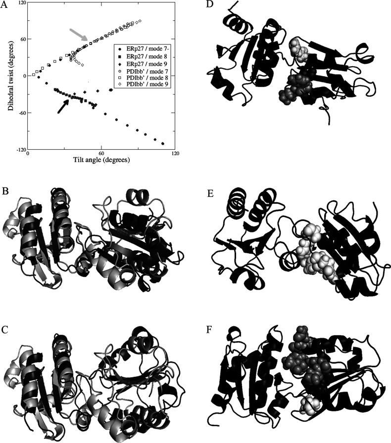
Figure 6. Domain orientation, flexibility and interdomain contacts in ERp27.
(A) Relative orientation of the β-sheet planes in adjacent Trx-fold domains for ERp27 and for the b-b′ moiety of yeast PDI. The planes are defined by the positions of four Cα atoms in each domain, as follows: for ERp27 domain b, Val60, Gly62, Ile106 and Leu108; for ERp27 domain b′, Leu164, Leu166, Leu222 and Ile224; for PDI b domain, Ile163, Gln165, Leu202 and Ile204; and for PDI b′ domain, Gly260, Leu262, Phe314 and Ile316. Orientation is described by a tilt angle between the plane normals, and by a dihedral twist angle formed by the plane normals and the interplane vector. The orientation in the PDB code 2B5E crystal structure of PDI is indicated by a grey arrow and that in the ERp27 crystal structure (PDB code 4F9Z [43]) by a black arrow. Symbols (open for PDI, closed for ERp27) show flexible motion along the three lowest-frequency non-trivial elastic network modes (modes 7, 8 and 9), in positive and negative directions, for each structure. Motion of ERp27 along mode 7− leads to PDI-like interdomain orientations. (B) Backbone cartoon overlay of ERp27 (black) with PDI b-b′ (white) crystal structures, aligned on the b′ domain (left-hand side) only, showing very different b-b′ orientations. (C) Corresponding backbone cartoon overlay of ERp27 after projection along mode 7− (black) with PDI b-b′ (white) crystal structure; the b domain β-sheets of ERp27 and PDI are now coplanar. (D) Backbone cartoon of ERp27 after projection along mode 7− (black). Residues 59 (dark grey), 82, 83 and 86 (all light grey), and 110 (white) are shown as spheres. These residues make new contacts with domain b′, not found in the input crystal structure. (E) Backbone cartoon of ERp27 after projection along mode 7+ (black), showing residues 110, 116, 117 and 118 as white spheres, forming new contacts with domain b′. (F) Backbone cartoon of ERp27 after projection along mode 8− (black), showing residues 59 and 60 (dark grey spheres), 80–83, 86 and 87 (light-grey spheres), and 110 (white spheres) forming new contacts with domain b′.

