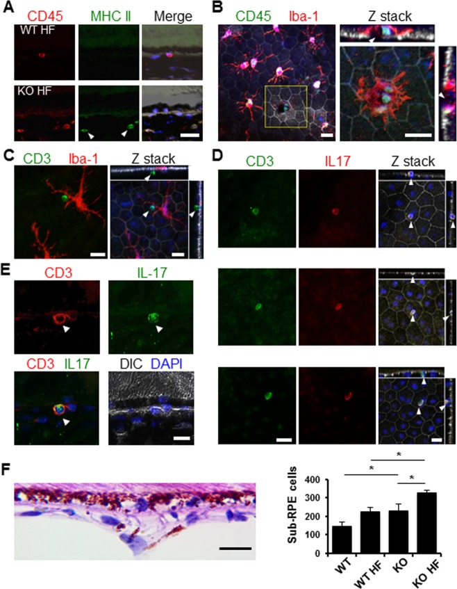
Figure 4.
Infiltration of IL-17-producing lymphocytes in the RPE/choroid complex of Nrf2-deficient mice on the HF diet. (A) Immunostaining of cryosections for MHC II and CD45. CD45 and MHC II double-positive cells (arrowheads) were mostly observed in Nrf2 knockout mice. (B–D) Immunostaining of RPE flat mounts from Nrf2−/− mice for lymphocyte markers. (B) CD45+ cells observed close to Iba-1+ microglia. Reconstruction of Z stack of the yellow boxed area showed CD45+ cells near the RPE defect as indicated by the discontinued tight junction (arrowheads in sidebars), surrounded by a microglia cell (red signal). (C) Adjacent CD3+ lymphocytes (arrowheads) and Iba-1+ microglia (red) separated by the RPE. (D, E) CD3 and IL-17 staining of RPE flat mount and cryosections, respectively. IL-17-producing lymphocytes (arrowheads) were identified on apical, in middle, or on basal side of the RPE layer (D). (F) Quantification of sub-RPE cells within 60-μm distance to the BrM on histology slides, showing significant increase of cells in knockout HF group compared to either normal chow–fed Nrf2−/− mice or wild-type mice on HF diet (*P < 0.05, one-way ANOVA and Bonferroni post hoc test). Scale bars: 20 μm.

