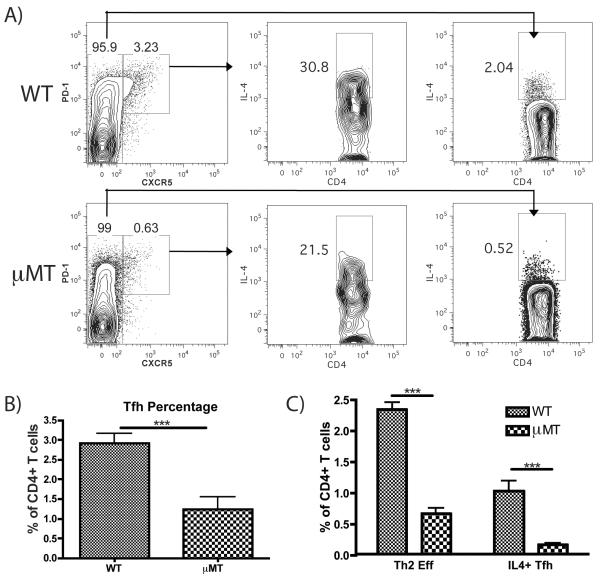
FIGURE 4.
μMT mice show impaired Tfh generation and reduced IL-4 induction by both Th2 and Tfh cells. (A) Representative FACS plots showing the induction of Tfh (PD1hi, CXCR5hi) and IL-4 expression by Tfh and conventional T cells (CXCR5lo) in WT and μMT mice immunized in the footpad with papain 5 d earlier. (B) Quantification of total Tfh as a percentage of all CD4+ T cells in WT and μMT mice on d 5 post-papain immunization. Bars indicates mean ± SD from 2 independent experiments with n=3 mice per group per experiment, *** indicates p<0.001. (C) Comparison of effector Th2 cells (IL-4+, CXCR5 low) versus IL-4+ Tfh cells (CXCR5hi, PD-1hi, IL-4+) as a mean percentage of all CD4+ T cells in WT and μMT mice 5 d post-immunization, calculated by multiplying the percentage of CD4+ T cells represented by CD4+ effector or Tfh cells (left panels in A) by the fraction of each population expressing IL-4 (center and right panels in A). Bars indicate mean ± SD from 2 independent experiments with n=3 mice per group per experiment, *** indicates p<0.001.

