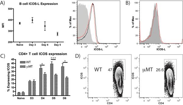
FIGURE 6.
PLN B cells express ICOS-L and μMT mice show impaired ICOS upregulation on PLN CD4+ T cell after papain immunization. (A) Time course of B cell ICOS-L MFI in the PLN of naïve mice and papain-immunized mice 3–5 d post-immunization and representative histogram indicating B cell ICOS-L expression in naïve animals (black) and in mice immunization with papain 5 d prior (red) compared to isotype control staining (grey). Points indicate mean ± SD from 2 independent experiments, n=3 mice per group per experiment. ANOVA p<0.05. (B) Representative histogram indicating ICOS-L expression on MHC-II+ CD11c+ DC in mice immunized with papain 5 d previously. Red indicates ICOS-L staining and grey indicates isotype control. (C) Time course showing the percentage of ICOS-expressing PLN CD4+ T cells in WT and μMT mice 3–6 d following papain immunization. Bars indicate mean ± SD from 2–3 experiments per time point with n=3 mice per group per experiment. * indicates p<0.05, *** indicates p<0.001 (D) Representative FACS plots of CD4+ T cell ICOS expression in WT and μMT animals 5 d post-immunization, indicating that ICOS upregulation occurs in a subset of T cells.

