FIGURE 7.
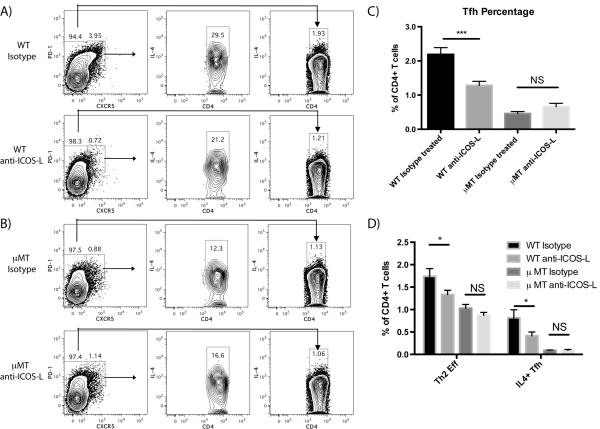
mAb blockade of ICOS-L markedly reduces Tfh expansion and IL-4 production by Th2 and Tfh cells following papain immunization in WT but not in μMT mice 5 d post-immunization. (A) Representative FACS plots showing the induction of Tfh cells (PD1hi, CXCR5hi) and IL-4 expression 5 d post-papain immunization by Tfh and conventional CD4+ T cells (CXCR5lo) in WT mice injected i.p. with either isotype control antibody or anti-ICOS-L mAB on d 0. (B) Representative FACS plots showing the induction of Tfh and IL-4 expression 5 d post-papain immunization by Tfh and conventional T cells in μMT mice injected i.p. with either isotype control antibody or anti-ICOS-L mAb on d 0. (C) Quantification of total Tfh as a percentage of all CD4+ T cells 5 d post-papain immunization in WT and μMT mice injected with either isotype control antibody or anti-ICOS-L. Bars indicate mean ± SD of n=2–3 independent experiments with 3–4 mice per experiment, *** indicates P<0.001, NS indicates no significant difference. (D) Comparison of effector Th2 cells (IL-4+, CXCR5lo) vs IL-4+ Tfh cells (CXCR5hi, PD-1hi, IL-4+) as a mean percentage of all CD4+ T cells 5 d post-papain immunization in WT mice injected with either isotype control antibody or anti-ICOS-L. Bars indicate mean ± SD of n=2–3 independent experiments with 3–4 mice per experiment, * indicates P<0.05.

