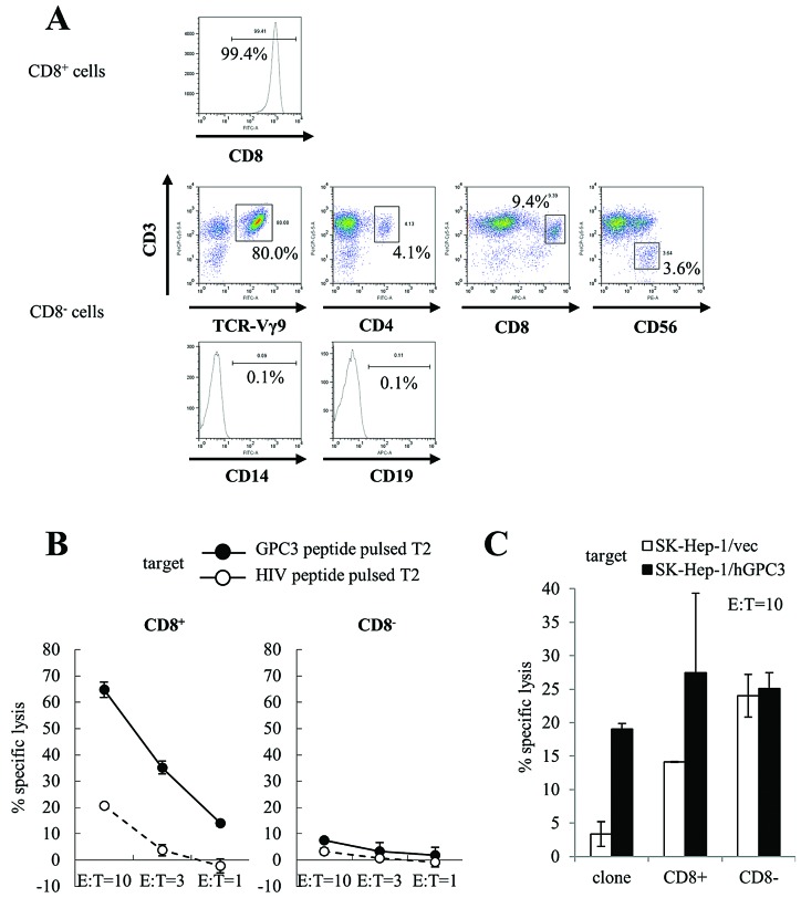
Figure 4.
Cytotoxicity assay of cultured cells. We used CD8+ and CD8− cells that were isolated from cultured cells using CD8 microbeads at day 14 as effector cells. A GPC3 peptide-specific CTL clone was used as a positive control. We performed cytotoxicity assays using expanded cells from four patients. Similar results were obtained in three of the four patients. Representative data are shown. (A) We examined the purity of the CD8+ cell populations obtained for these experiments. We performed further immunophenotyping of CD8− cells using flow cytometry. (B) T2 cells pulsed with GPC3 (black circle) or HIV (white circle) peptide were used as target cells. CD8+ cells (left) showed GPC3 peptide-specific cytotoxic activity. CD8− cells (right) did not show GPC3 peptide-specific cytotoxic activity. (C) SK-Hep-1/hGPC3 (black bar) or SK-Hep-1/vec (white bar) cells were used as target cells. CD8+ cells showed GPC3-specific cytotoxicity, whereas CD8− cells showed cytotoxicity against SK-Hep-1 cells, but not GPC3 specificity (E:T=10). Data represent the means ± SD.

